

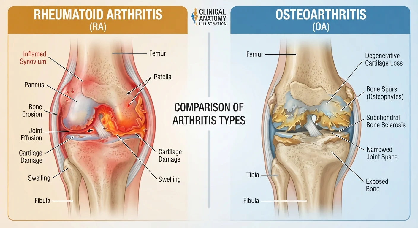
Option A
Rheumatoid Arthritis (RA)
An autoimmune disease that attacks the joint lining.
Best for: Understanding immune-driven joint inflammation affecting people of any age, including younger adults.
Option B
Osteoarthritis (OA)
A mechanical, wear-related breakdown of joint cartilage.
Best for: Understanding age- and use-related cartilage deterioration, most common in adults over 50.
Fundamentally Different Diseases With a Similar Symptom
Joint pain is the shared surface between rheumatoid arthritis (RA) and osteoarthritis (OA), but the similarity largely ends there. RA is an autoimmune disease — the immune system mistakenly targets the synovium, the tissue lining the joints, triggering chronic inflammation that can damage cartilage, bone, and surrounding structures. OA, by contrast, is a degenerative joint condition driven by the gradual breakdown of cartilage, the cushioning tissue that prevents bones from rubbing together.
This distinction in origin matters enormously for treatment. A person managing RA needs medications that modulate immune activity; a person managing OA needs strategies aimed at reducing mechanical load and managing pain. Treating one like the other can delay effective care.
For a broader look at how arthritis differs from bone-loss conditions, see our comparison of arthritis and osteoporosis.
| Criterion | Rheumatoid Arthritis (RA) | Osteoarthritis (OA) |
|---|---|---|
| Underlying cause | Autoimmune — immune attacks synovium | Degenerative — cartilage wear and breakdown |
| Typical age of onset | 30–60 years (can occur at any age) | Usually 50+ years |
| Joint pattern | Symmetrical (both sides equally) | Asymmetrical (one side may be worse) |
| Morning stiffness | Prolonged — often 1+ hour | Brief — usually under 30 minutes |
| Systemic symptoms | Fatigue, fever, weight loss common | Generally absent |
| Key diagnostic tests | RF, anti-CCP antibodies, CRP, ESR | X-ray findings; blood work usually normal |
| Primary treatment goal | Suppress immune activity; prevent erosion | Relieve pain; preserve function |
| Joints most affected | Hands, wrists, feet, knees | Knees, hips, hands, spine |
Symptoms, Onset, and Who Is Most Affected
RA most commonly develops between ages 30 and 60, and it affects women at roughly two to three times the rate of men, according to the American College of Rheumatology. Its onset is often gradual, beginning with symmetrical joint swelling — the same joints on both sides of the body — along with warmth, redness, and pronounced morning stiffness lasting an hour or more. Systemic effects including fatigue, low-grade fever, and loss of appetite are common because RA is a whole-body inflammatory condition.
OA tends to develop after age 50, though it can occur earlier following joint injury. It typically involves asymmetrical joint involvement, meaning it often affects one knee or hip more than the other. Pain characteristically worsens with activity and eases with rest. Morning stiffness in OA is usually brief — generally under 30 minutes — and there are no systemic symptoms.
1.3M+
Americans estimated to have RA
The American College of Rheumatology estimates over 1.3 million U.S. adults are affected by rheumatoid arthritis.
32.5M
U.S. adults affected by osteoarthritis
The CDC reports approximately 32.5 million U.S. adults have osteoarthritis, making it the most common form of arthritis.
2–3×
Higher RA prevalence in women vs. men
Women are diagnosed with RA at two to three times the rate of men, according to American College of Rheumatology data.
Small joints of the hands, wrists, and feet are frequent early targets in RA. OA most commonly affects the knees, hips, hands (particularly the base of the thumb and finger-tip joints), and the spine.
Diagnosis: Why Blood Tests and Imaging Matter
No single test confirms either condition, but the diagnostic pathways differ. For suspected RA, clinicians typically order blood tests for rheumatoid factor (RF) and anti-cyclic citrullinated peptide (anti-CCP) antibodies, along with markers of inflammation such as C-reactive protein (CRP) and erythrocyte sedimentation rate (ESR). Imaging — X-rays and increasingly MRI — helps assess joint erosion. Importantly, some people with RA test negative for RF (seronegative RA), so clinical judgment remains central.
Seronegative RA: When Blood Tests Are Negative
Up to 20–30% of people with RA test negative for rheumatoid factor (seronegative RA). Anti-CCP antibodies are more specific and may still be positive, but some patients test negative for both markers. In these cases, diagnosis depends on clinical symptoms, imaging findings, and specialist evaluation. A negative blood test does not rule out RA.
OA diagnosis relies more heavily on a person's symptoms, physical examination findings, and characteristic X-ray changes such as joint space narrowing, bone spurs (osteophytes), and subchondral bone changes. Blood markers are generally normal in OA.
Both conditions require a clinician's evaluation for accurate diagnosis. Attempting to self-diagnose based on symptoms alone is unreliable and can lead to inappropriate self-management.
Treatment Approaches and Long-Term Management
RA treatment has been transformed by disease-modifying antirheumatic drugs (DMARDs), including conventional agents and newer biologic therapies. The goal is to suppress the immune attack, prevent joint erosion, and achieve remission where possible. Early, aggressive treatment is associated with better long-term outcomes. Nonsteroidal anti-inflammatory drugs (NSAIDs) and corticosteroids may be used short-term to control flares but are not sufficient as standalone therapies for RA.
OA management is primarily focused on symptom relief and functional preservation. Physical therapy, weight management, and low-impact exercise are cornerstones of care. Pain relief may involve topical or oral analgesics, and for appropriate candidates, joint injections or surgical options such as joint replacement may be considered. For a detailed look at how lifestyle and clinical interventions compare, see our guide to osteoarthritis management.
Across both conditions, appropriate physical activity remains important — but the type, intensity, and precautions differ. Always discuss an exercise plan with your healthcare team before starting.
This article is for general informational purposes only and does not constitute medical advice. If you are experiencing joint pain, stiffness, or other symptoms, consult a qualified healthcare professional for evaluation and guidance.
The content on this site is for informational purposes only and is not a substitute for professional advice. Always consult a qualified professional for guidance specific to your situation.